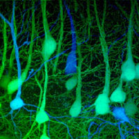
Take a genetically-engineered mouse and add color. That is what Jeffrey Lichtman, Jean Livet, and Joshua Sanes have done. Start by inserting genes that turn neurons fluorescent hues of yellow, red and cyan. Then add some more DNA that randomly expresses those three genes. Presto, rainbow brains.

As a Harvard Science piece reports, “By activating multiple fluorescent proteins in neurons, neuroscientists at Harvard University are imaging the brain and nervous system as never before, rendering their cells in a riotous spray of colors dubbed a ‘Brainbow.’ This technique… allows researchers to tag neurons with roughly 90 distinct colors, a huge leap over the mere handful of shades possible with current fluorescent labeling.”
So many colors in something as complex and elegant as a neuron produces striking images, and I have included many here. These images also permit the study of fields of neurons, from the life course of one neuron to the patterns of connections between neurons. Hence the emerging field of “Connectomics” which “attempts to physically map the tangle of neural circuits that collect, process, and archive information in the nervous system.”

I stumbled across Lichtman’s images in two publications recently. Harvard Magazine features his work, along with five other Harvard scientists, in this month’s feature article, Shedding Light on Life: Advances in Optical Microscopy Reveal Biological Processes as They Unfold. The magazine also provides an online collection of short video clips called Lights! Microscopes! Action! Across the Charles River, MIT’s Technology Review features Lichtman’s work as one of its Ten Emerging Technologies of 2008, complete with an accompanying video featuring Lichtman.
How does the wiring of the brain work? What happens when things go awry? The combination of new approaches to imaging, molecular biology, and computational analysis offer us up new maps. As Lichtman describes in the MIT video, he is like the explorers of old, discovering new lands never before seen. (Don’t worry, he also quotes Yogi Berra, “You can observe a lot by watching.”)

I am often as interested in ways of thinking as I am in specific results on problems. Faced with life’s diversity, styles of thinking—our core assumptions and analytical approaches—shape both results and interpretations. Lichtman’s style works.
Take this crappy clip on YouTube, lifted from a National Geographic program. Lichtman tells us, “We don’t normally think of nerve cells as living organisms, but in fact, they are alive. They just happen to be living inside your brain. But they all have to survive, and to do so, they have to compete with each other for resources.”

Cells as living organisms—a very different metaphor than machines or modules in our brain. Moreover, Lichtman approaches the study of nerve cells as a naturalist. In this Harvard press release, his student Thomas Misgeld tells us, “We are naturalists, trying to visualize processes that have so far escaped observation.” The piece goes on, “While most researchers use microscopes to ‘count and quantify’ the results of something they’ve done, the Lichtman team uses them to explore the inner life of the nervous system much as the Hubble telescope is used to probe space. ‘This work requires skills that you have to learn over time. Jeff is very good at identifying details in images that you thought were odd, but you hadn’t quite picked up on them,’ Misgeld says.”
As a good biologist, Lichtman also draws on evolutionary theory, in particular taking a Darwinian stance on neuronal selection. Here’s a good example from a Harvard Gazette piece that covers his work on development and the nerve innervation of muscle tissue:
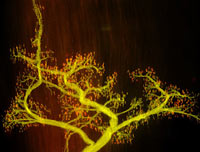
There’s a war going on inside our bodies, early in life. The combatants are motor nerve cells, the strangely branched bodies that carry nerve signals from our brains to our muscles and that are responsible for all our movements, from a sprint across a field to the tiniest twitch of a finger. The prize is the muscle fibers they are struggling to control. Each nerve cell sends out multiply-branched fingers called axons that contact many muscle fibers, which are also in contact with axons from other nerve cells.
And a battle ensues. The different nerve endings compete back and forth until one is the victor and becomes the conduit for messages from the brain to that particular bit of muscle. In the meantime, the battle leaves us pretty helpless.
That, according to new Professor of Molecular and Cellular Biology Jeff Lichtman, is the real reason human babies start out as needy as they do in life. “A baby can’t turn over, can’t understand language, its coordination is lousy, it can’t see very well,” Lichtman said. “There’s something not right about every aspect of their nervous system.”
But Lichtman does not use an innatist Darwinian approach. As we have argued many times here, the ways our brains are wired (and thus function) are activity- and experience-dependent. In a 2003 Letter to Nature, the Lichtman/Sanes group argues: “Synaptic activity drives synaptic rearrangement in the vertebrate nervous system; indeed, this appears to be a main way in which experience shapes neural connectivity. One rearrangement that occurs in many parts of the nervous system during early postnatal life is a competitive process called ‘synapse elimination’… In support of the idea that synapse elimination is activity dependent, it is slowed or speeded when total neuromuscular activity is decreased or increased, respectively… Here we use a genetic method to selectively inhibit neurotransmission from one of two inputs to a single target cell. We show that more powerful inputs are strongly favoured competitors during synapse elimination.”

The same group then backs that up by also considering the specific mechanism behind the elimination process. In a 2004 Neuron article, the authors argue, “Synapse elimination has been extensively studied at the neuromuscular junction, but how axons are lost is unknown. Here, we combine time-lapse imaging of fluorescently labeled axons and serial electron microscopy to show that axons at neuromuscular junctions are removed by an unusual cellular mechanism. As axons disappear, they shed numerous membrane bound remnants. These ‘axosomes’ contain a high density of synaptic organelles and are formed by engulfment of axon tips by Schwann cells. After this engulfment, the axosome’s contents mix with the cytoplasm of the glial cell. Axosome shedding might underlie other forms of axon loss and may provide a pathway for interactions between axons and glia.” The Lichtman Lab also provides webvideo and stills of this work, in case you want to see it in action.
For a final element in his approach to problems, Jeff Lichtman is interested in connections and networks, not only mechanisms and selection or how experience drives neuronal function. As a National Geographic News piece tells us, “Genetically engineered mice furnished with fluorescent proteins are providing the most detailed pictures yet of the brain’s intricate circuitry… ‘Imagine the brain as a radio for which we never had a good wiring diagram,” said Jeff Lichtman, a neurobiologist at Harvard University and a co-author of the study. ‘The aim of this work is to tag the individual wires with their own color’ to get a better idea of their connections’.”
Why is this important? In a general sense, biology and anthropology alike are trapped in analyzing problems from either an individual or a population-level. We do not understand enough about the dynamics that link individuals, whether neurons or people, into complex and functional patterns. Social network analysis and connectomics offers a way to tackle this sort of problem.
As the MIT Technology Review article says: “Lichtman and others hope that the ability to study multiple neural circuits simultaneously and in depth will provide unprecedented insight into how the wiring of the nervous system can go awry. ‘There’s a whole class of disorders of the nervous system that people suspect are due to defects in the connections between nerve cells, but we don’t have real tools to trace the connections,’ says Lichtman. ‘It would be very useful to look at wiring in animal models of autism-spectrum disorders or psychiatric illness’.”
To sum up, besides the synergistic research in itself and the accompanying beauty of the photographs, Lichtman’s style of thinking sets his approach apart—biology based in both a naturalist and a Darwinian framework, coupled with a concern with experience-dependent phenomenon, underlying mechanisms, and the role of networks. Sounds like a good way to think about life.
Image Credits: The photos here appeared in the articles cited in this post, with specific attributions to Jean Livet, Tamily Weissmann, and the Lichtman Lab. To see more, check out this Harvard Science Somewhere Inside the Brainbow slideshow. I also have a follow-up post, More on Brainbow, which answers questions about how the researchers get those flourescent colors and the pictures too, as well as some more images and even a short video.

For those of you interested in even more images, Wired has an article “Neurons or Pollock? Scientists Create Technicolor ‘Brainbow’.” There are another six images there that don’t appear above: http://www.wired.com/science/discoveries/multimedia/2007/10/gallery_fluorescentneurons
Why does Jeff Lichtman prefer his approach to biology, even though it is not common in his field of study?
His peices are fascinating through-out! Pondering upon neurons often leads me to the greatest example nature can offer, a tree. Age, gives us the chance to grow, to become strong and rooted in this world, and i love how Jeff captues this. Beautiful.
Peace ~